Figure 4.
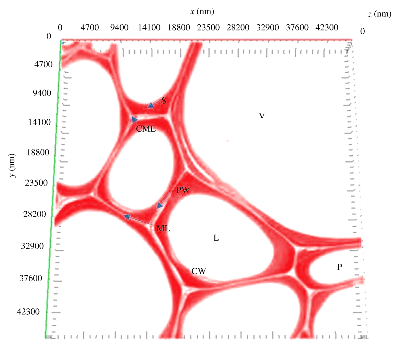
‘Confocal microscopy showing the cross-section cell wall structure with autofluorescence. V, vessel; S, second wall; P, parenchyma; L, lumen; PW, primary wall; ML, middle lamella; CML, compound middle lamella; CW, cell wall.‘
In: Jiang, Y., Lawrence, M., Ansell, M. P., & Hussain, A. (2018). Cell wall microstructure, pore size distribution and absolute density of hemp shiv. Royal Society Open Science, 5(4), 171945, p.7