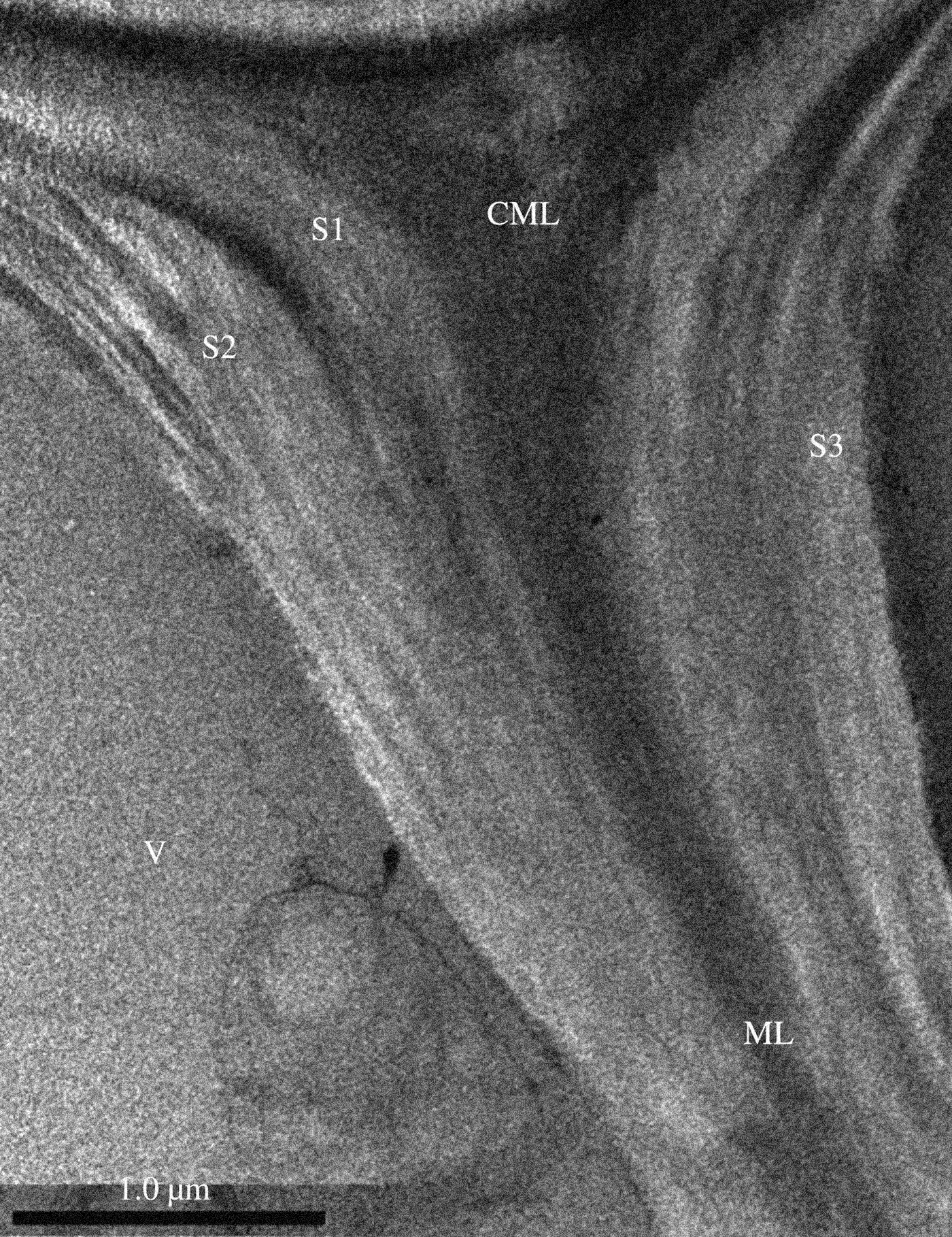
Figure 5.
‘TEM micrographs of cross-section cell wall structure. V, vessel; S1, outer secondary wall of vessel; S2, middle secondary wall of vessel; S3, inner secondary wall of vessel; ML, middle lamella; CML, compound middle lamella.’
In: Jiang, Y., Lawrence, M., Ansell, M. P., & Hussain, A. (2018). Cell wall microstructure, pore size distribution and absolute density of hemp shiv. Royal Society Open Science, 5(4), 171945, p.8