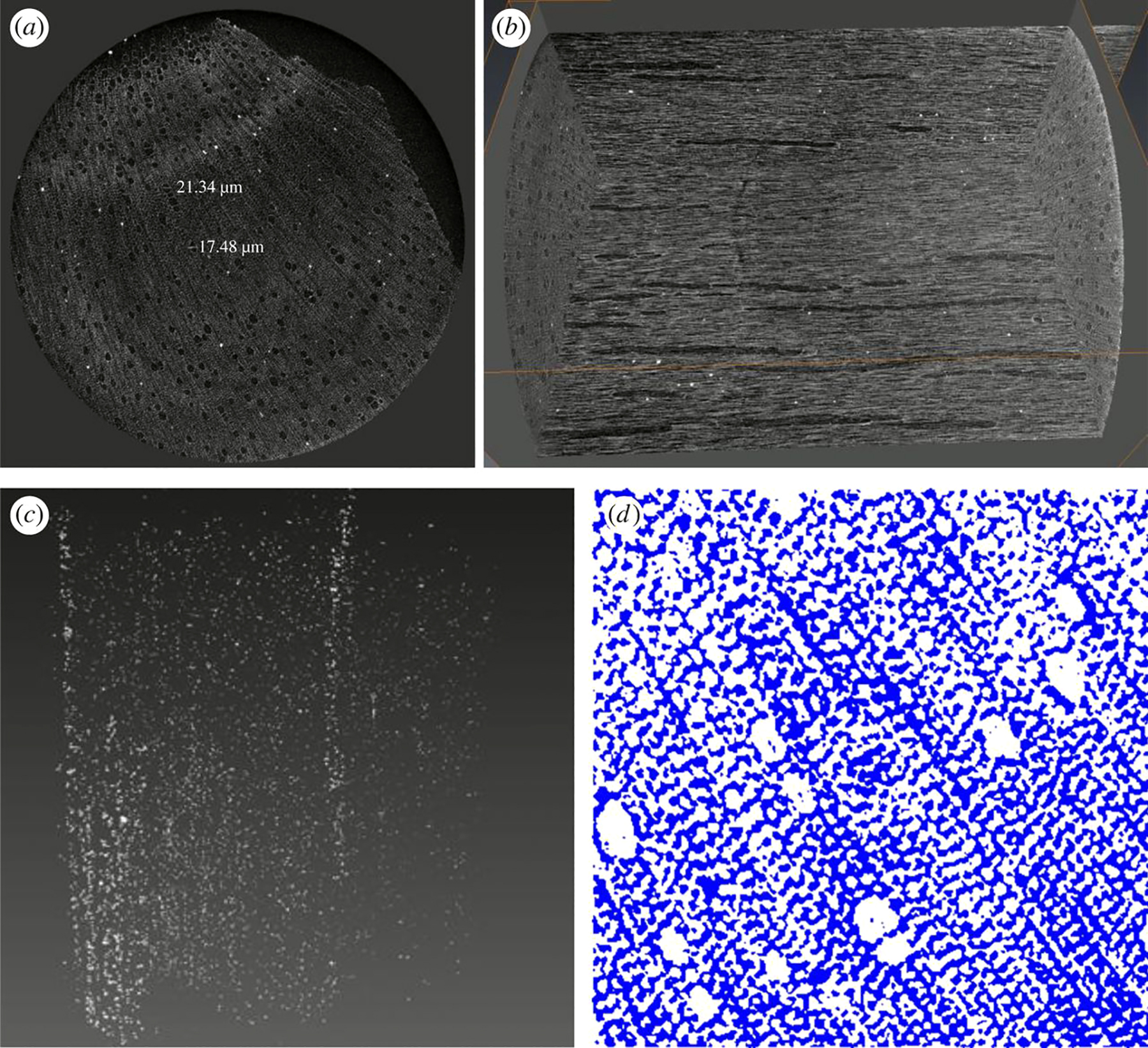
Figure 6.
‘CT scanning measurement data: (a) volume rendering of a hemp shiv specimen cross section, (b) internal structure, (c) distribution of warts and (d) cross-section image segmented with the global LA-Kriging method (threshold 125). White is air pore and blue is solid.’
In: Jiang, Y., Lawrence, M., Ansell, M. P., & Hussain, A. (2018). Cell wall microstructure, pore size distribution and absolute density of hemp shiv. Royal Society Open Science, 5(4), 171945, p.9